
Cerebrospinal fluid (CSF) Analysis
What is CSF ?
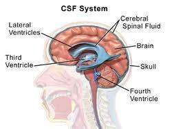
The Cerebrospinal fluid (CSF) is formed by selective dialysis of plasma by the choroid plexus of the ventricles of the brain . Through the foramina in tha fourth ventricle it then passes into subarachnoid cisterns at the base of the brain and travels over the surface of the cerebral hemispheres . It is finally absorbed into the blood in the cavity that surrounds the brain in tha skull and the spinal cord in the spinal column . The volume of CSF ( adult ) is about 150 ml .
CSF performed following functions :-
- It helps to protect the brain and spinal cord from injury by acting like a fluid buffer .
- It also acts as a medium for the transfer of substance from the brain tissue and spinal cord to blood .
- It maintains intracranial pressure .
Normal composition of CSF :-
| color | coloriess |
| pH | 7.3 – 7.4 जेड |
| Appearance | clear |
| clot formation | No clot formation on standing |
| specific gravity | 1.003 – 1.008 |
| Total solids | 0.85 – 1.70 g/dl |
| Protein | 15 to 45 mg/dl ( albumin = 50 – 70 % ) ( globulin = 30 – 50 % ) |
| Glucose | 40 – 80 mg/dl |
| cholorides | 700 – 750 mg/dl |
| sodium | 144 – 154 mEq/l |
| Potassium | 2.0 – 3.5 mEq/l |
| Creatinine | 0.5 – 1.2 mg/dl |
| cholesterol | 0.2 – 0.6 mg/dl |
| urea | 6 – 16 g/dl |
| uric acid | 0.5 – 4.5 mg/dl |
| cells | 0 – 8 Lymphocytes/per Cumm ( microlitter) ( Neutrophils absent) |
Clinical significance :-
CSF examination is carried out in the laboratory mainly for the diagnosis of meningitis . It is inflammation of the meaninges the lining of the skull and covering of the brain and spinal column . Meningitis cause disturbance in the central nervous system . The other clinical conditions in which CSF examination may be required are encephalitis subarachnoid hemorrhage spinal cord tumor multiple sclerosis central nervous system syphilis etc CSF examination is also carried out in the treatments of elevated CSF pressure in selected patients with benign intracranial hypertension .
Specimen collection :-
- The specimen should be collected by a physician a specially trained technicians or nurse .
- The sterlie lumber puncture needle is inserted between the 3rd and 4th lumber vertebra to a depth of 4 to 5 cm .
- After the withdrawal of style the fluid is collected through the needle into two test tubes .
- Tube 1 ( sterile tube ) About 0.5 ml or few drops of CSF
- Tube 2 About 3 to 5 ml of CSF .
Important Precautions :-
- The collected CSF specimen must be examined immediately ( at least within one hour of the collection ) .
- The specimen collected for bacterial culture should not be stored in the refrigerator . ( The commonly sought pathogens Neisseria meningitisidis is killed by exposure to cold ) . The specimen ment for biochemical test only may be stored at 2 – 8 °C for 2 to 3 hours .
- Cells and trypanosomes are rapidly lysed after the collection of CSF . Hence urgent analysis of CSF is necessary .
- The specimen is difficult to collect hence once it is collected it is necessary to analyse the specimen carefully and economically .
- The specimen may contain virulent orgnisam hence it is necessary to handle it carefully .
Routine Examination of CSF :-
It is carried out by :-
- physical examination .
- microscopic examination .
- chemical examination .
physical Examination of CSF : –
observe the specimen and note down observations for the following aspects :
- color
- Appearance
- presence of blood
- presence of clot or fibrin web
use pH paper ( range 2 to 10.5 ) to determine pH .
If necessary determine specific gravity by the weight method ( weight of CSF/ volume of CSF ) or by using a hand refrectometer .
Microscopic Examination of CSF : –
Requirements :-
- glass slides
- Fuchs-Rosenthel counting chamber or improve Neubauer counting with coverslip .
- pasteur pipettes
- Leishman stain and buffer solution pH 7.0
- CSF diluting fluid
- grams staining reagents
- Acidfast staining reagents
- Centrifuge machine
- Microscopic
- Nichrome loop and bunsen burner
Stain Bacteria In Culture By Grams Staining method
Reagents
- crystal violet stain
solution A :-
crystal violet = 2 g
Ethyl alcohol = 20 ml
solution B :-
Ammonium oxalate = 0.8 g
Distilled water = 80 ml
Mix solution A and B . keep for 24 hours and filter . Store in an amber colored dropping bottle .
- Grams iodine solution
Iodine = 1.0 g
potassium iodide = 2.0 g
Distilled water = 100 ml
store in amber colored dropping bottle .
- Decolorizer
mix 95 % alcohol and acetone in equal proportion . ( If alcohol is not available rectified spirit can be used . ) Store in a white dropping bottle .
- safranin solution
safranin o = 0.34 g
absoulte alcohol = 10 ml
Distilled water = 90 ml
fillter and Store in an amber colored dropping bottle .
stability of the reagents
All the reagents are stable at room temperature ( 25°C +_ 5°C ) .
specimen :-
cultured cerebrospinal fluid .
Procedure : –
Smear preparation
- Take a gress free slide and make an oval shaped mark at the centre by using a glass marker .
- sterilize the inoculating ( nichrome ) loop on the flame of a burner .
- Transfer a loopful of culture ( or specimen ) by the sterile nichrome loop and marke a smear in the premarked area on the slide ( smear should not be very thin as well as very thick ) .
- Allow the smear to dry in the air .
- Fix the dry smear by passing the slide 3 to 4 times through the flame quickly with the smear side facing up .
Gram staining
- place the slide on the Staining glass rods .
- cover the smear with crystal violet stain and leave for 1 minutes .
- wash carefully under running tap water .
- floos the smear with grams iodine solution and wait for one minute .
- drain off the iodine .
- Decolorize the smear with alcohol acetone ( or rectified spirit ) for 20 to 30 seconds . Continue till purple stain just stops coming on the slide .
- Gently wash the slide under running tap water and drain completely .
- Counterstain the smear with safranin for 10 seconds or with dilute ( 1:20 ) basic Fuchsin for about 1 minutes .
- Drain the staining solution and allow the stained smear to dry in air ( or dry it carefully by using a blotting paper ) .
- First observe for uniform stained area under low power objective afterwards under high power objective and finally under oil immersion objective .
Physical Examination of Urine.
Precautions : –
It is necessary to standardize the time for steps 5 and 6 variation in these timings may lead to false results .
Results : –
The Staining results of gram stain are as follows
- fram negetive bacteria = pale to dark red
- gram positive bacteria = Dark purple
- yeast cells = Dark purple
- Nuclei of pus cells = red
- Epithelial cells = pale red
Total Leukocytes count
- mix the CSF sample carefully .
- Fill the Fuchs-Rosenthel chamber or Neubauer chamber with the CSF sample .
If CSF appears clear use it undiluted .
If CSF appears cloudy make 1:20 dilution by using a WBC pipette ( draw CSF specimen up to 0.5 mark and dilute with the diluting fluid up to 0.5 Mark and dilute with the diluting fluid upto 11 Mark mix well ) or pipette 9.95 ml of CSF diluting fluid in a small bottle and add 0.05 ml of CSF into it . Mix well .
- Leave the counting chamber on the counter for 5 minutes to allow the cells to settle .
- place the chamber on the microscope stage .
- Count the cells in 5 square ( using squares 1 , 4 ,7, 13, and 16 ) by using × 10 objective ( area counted = 5 mm square ) .
Leuckocytes in CSF/per Cumm ( microlitter ) = Number of cells counted / Aare counted × depth of fluid
chemical Examination of CSF Qualitative of globulins protein by pandys test : –
clinical significance : –
This test gives an indication of the extent of any increase in globulin in bacterial infection general paralysis of the insane and in disseminated sclerosis .
pandys regent : –
It is prepared by dissolving 10 g of phenol in 150 ml of distilled water . It should be clear and colorless .
Procedure : –
- pipette 2ml of pandys reagent in a small test tube ( 100 × 75 ) .
- Add 2 to 3 drops of clear CSF specimen do not mix .
- observe for the formation of turbidity .
Observations : –
- No formation of precipitate = Globulin Normal
- Formation of precipitate ring = Globulin Increased