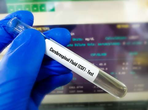
Cerebrospinal Fluid (CSF) Analysis
INTRODUCTION
The cerebrospinal Fluid (CSF) is formed by selective dialysis of plasma by the coroid plexus of the ventricle of the brain. For the foramina in the fourth ventricle it then passes into subarachnoidal cisterns at the base of the brain and travels over the surface of the cerebral hemispheres . It is finally absorbed into the blood in the cerebral veins and dural sinuses. CSF is present in the cavity that surrounds of the brain in the skull and in spinal cord in the spinal column. The volume of cerebrospinal Fluid (CSF) in adult is 150 ml.

FUNCTION OF CSF
Cerebrospinal Fluid (CSF) performing following functions.
- It help to protect the brain and spinal cord from injury by acting like a fluid buffer.
- It also act as a medium for the transfer of substance from the brain tissue and spinal cord to blood.
- It maintains intracranial pressure.
NORMAL COMPOSITION OF CSF
| Color | Colorless |
| pH | 7.3-7.4 |
| Appearance | Clear |
| Clot formation | Not clot formation on standing |
| Specific gravity | 1.003-1.008 |
| Total solids | 0.85-1.70 g/dl |
| Protein | 15-45 mg/dl |
| Albumin | 50%-70% |
| Globulin | 30%-50% |
| Glucose | 40-80mg/dl |
| Chlorides | 700-750 mg/dl |
| Sodium | 144-154 mEq/l |
| Potassium | 2.0-3.5 mEq/l |
| Creatinine | 0.5-1.2 mg/dl |
| cholesterol | 0.2-0.6 mg/dl |
| Urea | 6-16 g/dl |
| Uric acid | 0.5-4.5 mg/dl |
| Cells | 0-8 lymphocytes /cumm (neutrophils : absent) |
SPECIMEN COLLECTION
- The specimen should be collected by a physician, a specially trained technician or nurse.
- The sterile lumber puncher needle is inserted between the 3rd and 4th lumber vertebra to a depth of 4 to 5 cm.
- After the withdrawal the stylet the fluid is collected through the needle into 2 test tube.
- Tube 1 : (sterile tube) : About 0.5 ml or few drops of CSF.
- Tube 2 : About 3-5 ml of CSF.
- The specimen in tube 1 may be used for bacterial culture if necessary.
- Specimen in the second give is centrifuged.
- Supernatant is used for the biochemical test such as glucose, protein, globulin and chlorides.
- Use the sediment for the following purpose.
- Prepare 3 smears.
- Smear 1 : Grams staining
- Smear 2 : Acid- fast staining
- Smear 3 : Differential leukocyte count
- Prepare wet mount for Trypanosoma.
- Proceed for India ink preparation in case of Cryptococcus infection.
- Prepare 3 smears.
IMPORTANT PRECAUTIONS
- The collected CSF specimen must be examined immediately, (at least within one hour of the collection).
- The specimen collected for bacterial culture should not be stored in the refrigerator (The commonly sought pathogen Neisseria meningitidis is killed by exposure to cold) the specimen meant for biochemical test only, may stored at 2-8 degree Celsius for 2-3 hours.
- Cells and trypanosomes are rapidly lysed after the collection of CSF. Hence urgent analysis of CSF is necessary.
- The specimen is difficult to collect, hence once it is collected, it is necessary to analyse the specimen carefully and economically.
- The specimen may contain virulent organisms, hence it is necessary to handle it carefully.
CLINICAL SIGNIFICANCE
Cerebrospinal Fluid (CSF) Analysis is carried out in the laboratory mainly for the diagnosis of meningitis. It is inflammation of the meninges, the lining of the skull and covering of the brain and spinal column. Meningitis cause disturbance in the central nervous system. The other clinical conditions in which CSF examination may be required for encephalitis, subarachnoid hemorrhage, spinal cord tumour, multiple sclerosis, central nervous system syphilis, etc. CSF examination is also carried out in the treatment of elevated CSF pressure in selected patient with benign itntacranial hypertension.
ROUTINE EXAMINATION OF CSF
It is carried out by,
- Physical examination,
- Microscopic examination,
- Chemical examination
Physical Examination
- Observe the specimen and note down observations for the following aspects:
- Color
- Appearance
- Presence of blood
- Presence of clot or fibrin web
- Use pH paper (range 2 to 10.5) to determine pH.
- If necessary, determine specific gravity by the weight method (weight of CSF / volume of CSF) or by using a hand refractometer.
Microscopic Examination
- Requirements
- Glass slides
- Fuchs-Rosenthal counting chamber or improved Neubauer counting chamber with coverslip.
- Pasteur pipettes
- Leishman stain and buffer solution
- CSF diluting fluid
- Grams staining reagent
- Acid-fast staining reagent
- Centrifuge machine
- Microscope
- Total Leukocyte count
- Mix the CSF sample carefully.
- Fill the Fuchs-Rosenthal counting chamber or improved Neubauer counting chamber with the CSF sample.
- If CSF appears clear, use it undiluted.
- If CSF appears cloudy, make 1:20 dilution by using a WBC pipette (draw CSF specimen up to 0.5 mark and dilute with the diluting fluid up to 11 Mark mix well) or Pipette 9.95 ml of CSF diluting fluid in a small bottle and add 0.05 ml of CSF into it. Mix well.
- Leave the counting chamber on the counter for 5 minutes to allow the cells to settle.
- Place the chamber on the microscope stage.
- Count the cells in 5 square (using square 1,4,7,13 and 16) by using X10 objective (are counted = 5 mm2).
- Calculations: (by using Fuchs-Rosenthal counting chamber) Leucocyte in CSF / cumm = No. of cells counted / Area counted × depth of fluid
- Calculations: (if Neubauer counting chamber is used) Leucocyte in CSF / cumm = cells counted / 9 × 1/10
- Determination of differential leukocyte count = Click here
- Increased lymphocyte count is an indication of possible viral infection.
- Increased Neutrophils indicates bacterial infection.
Chemical Examination
Determination of globulin by Pandy’s test
- Clinical Significance : This test gives an indication of the extent of any increase in globulin and bacterial interaction, general paralysis of the insane and disseminated sclerosis.
- Pandys reagent : It is prepared by dissolving 10 gram of phenol in 150 ml of distilled water. It should be clear and colourless.
- Procedure :
- Pipette 2 ml of pandy’s reagent in a small test tube.
- Add 2 to 3 drop of clear CSF specimen. Do not mix.
- Observe for the formation of turbidity.
- Observations :
- No formation of precipitate : globulin : Normal.
- Formation of precipitate ring.
- Globulin : Increased
- Grade the positive results as trace, +, ++, +++, ++++, according to the degree of formation of the precipitate.
