
Preparation of Lupus Erythematosus (LE) Cell
INTRODUCTION
Antinuclear antibodies occur in the serum in a wide range of autoimmune disorders, including systematic lupus erythematosus (SLE). A characteristic of SLE is the presence of 7S IgG antibodies to double standard DNA (ds – DNA). LE (lupus erythematosus) cells are a type of abnormal immune cell that can be seen in the blood of some individuals with lupus erythematosus, a chronic autoimmune disease that can affect different parts of the body. LE cells are actually neutrophils (a type of white blood cell) that have engulfed and are carrying around a large, round structure known as an LE body.
LE cells are formed when antibodies that are produced by the immune system mistakenly attack and bind to the nucleus of the body’s own cells. This leads to the formation of immune complexes that are then engulfed by neutrophils, resulting in the formation of LE cells. Detection of LE cells in the blood can be helpful in the diagnosis of systemic lupus erythematosus, but it is not always present in all cases of lupus. In addition, there are other conditions where LE cells may be seen, such as viral infections and some other autoimmune diseases, so further diagnostic tests are usually required to confirm a diagnosis of lupus.
PRINCIPLE
The leucocytes are traumatized by rotating anticoagulated blood with glass bead, (since LE factor does not attack healthy living leucocytes). The patient’s serum containing LE factor i.e. antinuclear antibody (gamma globulin function) is then gets directed toward the nuclear material of leucocytes. The nuclear material is then transformed into an LE body which attacks the neutrophils and then ingested by one of them. A smear of Buffy coat is stained by using Leishman stain and examined under the high power Objective (X 40) in the microscope.
SPECIMEN
EDTA anticoagulated or clotted blood.
REQUIREMENTS
- Centrifuge tube, glass beads, rubber Bungs.
- Microscope glass slides, test tubes
- Leishman’s stain
- Centrifuge, microscope, blood mixing rotor or vortex mixer
PROCEDURE
- Take about 5 ml of EDTA blood in a test tube.
- Add 5 glass beads and stopper the tube.
- Rotate on a blood mixing router at 50 RPM for 30 minutes.
- Place the tube at 37 degree Celsius for 10 minutes.
- Transfer the blood to a wintrobe tube.
- Centrifuge wintrobe tube at 3000 rpm for 30 minutes.
- Remove hemolysed serum and then remove carefully the Buffy coat.
- Make smears of the Buffy coat on clean and dry glass slides.
- Fix the air dried smears in methanol.
- Stain by using Leishman’s staining solution.
- Screen under high Power (X 40) and observe specific areas under oil immersion objectives.
Erythrocyte Sedimentation Rate ( ESR ) Test
IMPORTANT NOTE
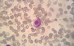
- LE cells appear as a neutrophil containing a large spherical bodies in its cytoplasm.
- The LE body does not show nuclear structure and stains as a pale purple homogenous mass.
- The ‘tart’ cells retain their nuclear structure and should not be confused with LE cells. A tart cell is a granulocyte that has engulfed the nucleus of another cell.
- Several LE cells should be identified before reporting the specimen to be positive.
- Other method involve reaction of patient’s serum with normal persons leucocytes. The procedure is exactly the same as mentioned above after mixing patients serum with normal leucocytes.
- If blood mixing rotor is not available, traumatization of white cells is carried out by continuous mixing of blood by rotating blood with 8 to 10 glass beads and a conical flask (50 ml) for 15 to 20 minutes.
- 7S IgG antibodies to double standard DNA(ds – DNA) can be detected by ELISA method.
IMPORTANT MCQS OF LE CELLS
What are LE cells?
- A) Lymphatic endothelial cells
- B) Lymphocyte-encapsulated cells
- C) Lupus erythematosus cells
- D) Lipid-encased cells
Answer: C) Lupus erythematosus cells
How are LE cells formed?
- A)When antibodies attack and bind to the nucleus of the body’s own cells, leading to the formation of immune complexes that are then engulfed by neutrophils
- B) When lymphocytes undergo mitosis and form a protective barrier around the body’s own cells
- C) When macrophages engulf and digest foreign pathogens
- D) When red blood cells are coated with antibodies and become agglutinated
Answer: A) When antibodies attack and bind to the nucleus of the body’s own cells, leading to the formation of immune complexes that are then engulfed by neutrophils.
Which condition is LE cell test useful for diagnosing?
- A) Rheumatoid arthritis
- B) Multiple sclerosis
- C) Lupus erythematosus
- D) Sickle cell anemia
Answer: C) Lupus erythematosus
In what type of lupus erythematosus are LE cells most commonly seen?
- A) Discoid lupus erythematosus
- B) Subacute cutaneous lupus erythematosus
- C) Systemic lupus erythematosus
- D) Neonatal lupus erythematosus
Answer: C) Systemic lupus erythematosus
Which type of white blood cell is most commonly associated with the formation of LE cells?
- A) Neutrophils
- B) Lymphocytes
- C) Monocytes
- D) Eosinophils
Answer: A) Neutrophils
What is the characteristic feature of an LE cell that distinguishes it from a normal neutrophil?
- A) A prominent nucleus with a visible nucleolus
- B) A prominent cytoplasmic granule
- C) The presence of an inclusion body within the cytoplasm
- D) The presence of a ring-like structure around the nucleus
Answer: D) The presence of a ring-like structure around the nucleus
Which antibody is most commonly associated with the formation of LE cells?
- A) Anti-Ro antibodies
- B) Anti-dsDNA antibodies
- C) Anti-Sm antibodies
- D) Anti-cardiolipin antibodies
Answer: B) Anti-dsDNA antibodies
Which of the following conditions is LE cell formation not commonly seen in?
- A) Viral infections
- B) Bacterial infections
- C) Fungal infections
- D) Parasitic infections
Answer: D) Parasitic infections
What is the appearance of an LE cell under a microscope?
- A) A normal neutrophil with an abnormally shaped nucleus
- B) A neutrophil with a ring-like structure around the nucleus containing an inclusion body
- C) A lymphocyte with an abnormally shaped nucleus
- D) A macrophage with a granular cytoplasm
Answer: B) A neutrophil with a ring-like structure around the nucleus containing an inclusion body
What is the diagnostic significance of the LE cell phenomenon?
- A) It is a specific marker for lupus erythematosus
- B) It is a sensitive marker for lupus erythematosus
- C) It is not specific or sensitive for lupus erythematosus
- D) It is a marker for other autoimmune diseases but not lupus erythematosus
Answer: C) It is not specific or sensitive for lupus erythematosus
What is the clinical significance of the presence of LE cells in the blood?
- A) It indicates active disease in lupus erythematosus
- B) It indicates a poor prognosis in lupus erythematosus
- C) It has no clinical significance in lupus erythematosus
- D) It is associated with an increased risk of infection in lupus erythematosus
Answer: A) It indicates active disease in lupus erythematosus
Which of the following drugs has been known to induce the formation of LE cells?
- A) Nonsteroidal anti-inflammatory drugs (NSAIDs)
- B) Penicillin
- C) Methotrexate
- D) Propranolol
Answer: D) Propranolol